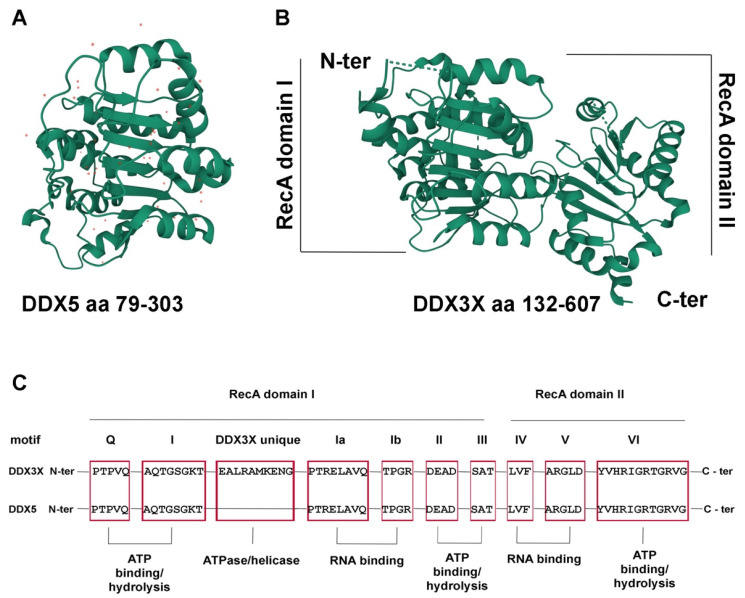
Figure 1.
The DDX3X and DDX5 structure. (A) Structure of the DDX5 N-terminal core domain (aa 79-303), PDB 4a4d [3]. Red dots represent water molecules. (B) Structure of the DDX3X helicase core (aa 132-607), PDB 5E7I [4]. The N-terminal domains of DDX5 and DDX3X are shown in the same orientation to highlight the conserved RecA fold of both proteins. Images were generated by the authors with the online tool Mol*3D viewer (https://www.rcsb.org/3d-view, accessed on 1 August 2022) [5]. (C) Schematic representation of the conserved helicase motifs of DDX3X and DDX5 (red boxes) with the respective functional roles.

