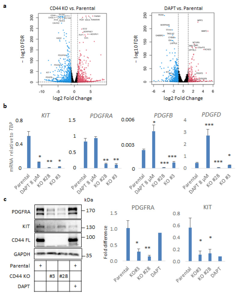
Figure 3.
CD44 ablation impairs autocrine PDGF signaling in U251MG cells grown in sphere-like conditions. (a) Volcano plots exhibiting differentially regulated genes, which were either down- or upregulated (depicted with blue and red color, respectively) in CD44 KO cells or parental U251MG cells treated with the γ-secretase inhibitor DAPT, compared to the untreated cells. (b) mRNA levels of KIT, PDGFRA, PDGFRB, and PDGFD determined by RT-qPCR in spheres formed by parental U251MG cells treated or not with DAPT, and CD44 KO cells, are shown after normalization to TBP. (c) Immunoblotting analysis of PDGFRA, KIT, and CD44 was performed using lysates from U251MG cells. GAPDH was used as the loading control. Uncropped immunoblots are depicted in Figure S9. Immunoreactive bands were quantified after normalization to GAPDH after using the software ImageJ. Cells were grown in low-attachment conditions in the presence or absence of the γ-secretase inhibitor DAPT. All graphs illustrate the average ± SEM values from at least three independent experiments. Asterisks show significant differences compared to the respective control condition: * p < 0.05, ** p < 0.01, *** p < 0.001.

