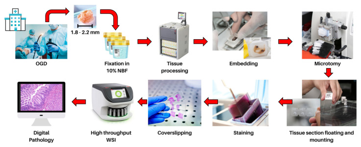
Figure 1.
Tissues obtained by endoscopic biopsies are fixed in 10% neutral buffered formalin for 12–24 h. The fixed tissue undergoes dehydration, clearing and impregnation with molten paraffin wax by automatic tissue processors. The tissue is subsequently embedded in a paraffin wax block with proper orientation so tissue sections (3–5 µm thick) can be cut with a microtome. The tissue section is manoeuvred onto a glass slide, stained and mounted with a coverslip to protect and preserve the section. The glass slide is digitized using high-throughput WSI scanners to create a virtual slide to allow for remote diagnosis and large-scale computational pathology.

