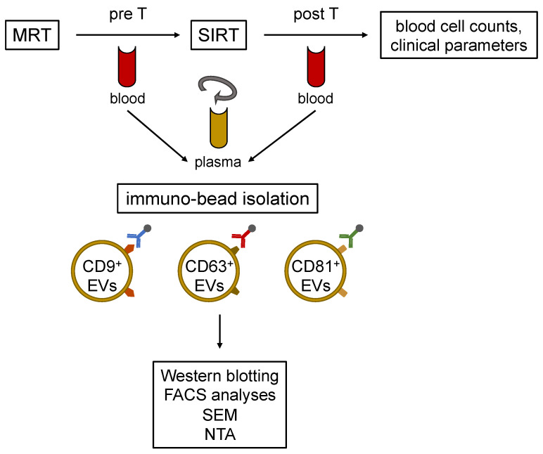
Figure 1.
Schematic illustration of the experimental workflow. Cholangiocellular carcinoma (CCA) patients received a liver MRI according to the clinical standards. Blood samples were taken before (pre T) and after (post T) selective internal radiation therapy (SIRT). Extracellular vesicles (EVs) were isolated from plasma using CD9+, CD63+, and CD81+ exosome markers in the immune-bead isolation. The isolated EVs populations are distinguishable by flow cytometry (FACS). The presence of EVs was assessed by western blotting, and in addition to the characterization of EVs, their morphology was analyzed by scanning electron microscopy (SEM) and nanoparticle tracking analysis (NTA).

