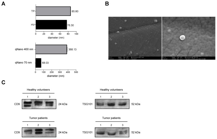
Figure 2.
Assessment of the quality of isolated extracellular vesicles (EVs) from healthy volunteers (HV) and tumor patients (TP) one day before (pre T) and two days (post T) after selective internal radiotherapy radioembolization. (A) Representative DLS- size distribution profile of calibration nanoparticles and isolated EVs collected in HV and TP. (B) Scanning electron microscopy image of of isolated EVs. The scale bar represents 2 µm, (10,000×; AccV. 10 kV) and 500 nm (35,000×; AccV. 12 kV) respectively. (C) Representative western blot assessing the protein content of isolated EVs from plasma of three different HV or TP (1–3) using genuine EVs markers CD9 and TSG101.

