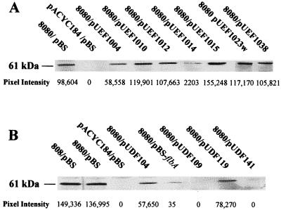
FIG. 4.
Western blot analysis of urease extracts by using anti-UreB. Cytosolic protein extracts (10 μg) were electrophoresed through an SDS-polyacrylamide gel, transferred to a PVDF membrane, and probed with anti-UreB as described in the Materials and Methods section. (A) Extracts from DH5α (pHP8080/pBluescript) and DH5α (pHP8080/pUEFs). (B) Extracts from SE5000 (pHP8080/pBluescript) and SE5000 (pHP8080/pUDFs). Density values are shown below individual blots. Results are representative of three experiments performed by using extracts prepared on 3 separate days. pBS, pBluescript; 808, pHP808; 8080, pHP8080.

