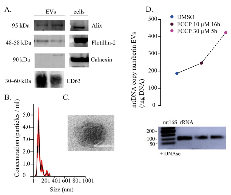
Figure 1.
Characterisation of human extracellular vesicles carrying mitochondrial DNA. (A) Western blots of human fibroblasts-derived EVs with traditional protein markers Alix, Flotillin-2 and CD63. Calnexin was used as a negative control for cell contamination. (B) Nanosight tracking analysis of EV concentration (particles/mL) and size (nm). (C) TEM image of the EV structure with visible lipidic bilayer. Scale: 100 nm. (D) Quantification of mtDNA copy number (/ng DNA) isolated from DNAse-treated EVs. Prior to EVs’ purification, cells were treated with 10 μM FCCP for 16 h and 30 μM FCCP for 5 h. DMSO was used as a control. PCR gel electrophoresis of mt16S isolated from DNAse-treated EVs. EVs: extracellular vesicles, TEM: transmission electron microscopy. Adapted from https://doi.org/10.1101/2022.02.13.480262; accessed on 3 June 2022.

