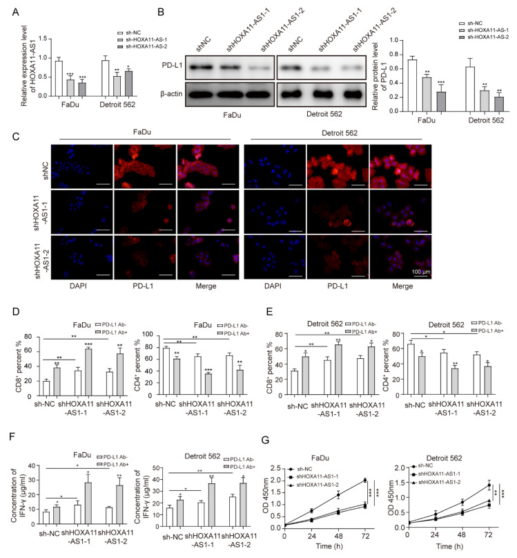
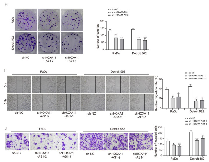
Figure 2.
HOXA11-AS1 knockdown suppressed PD-L1 expression and immune escape, proliferation, and metastasis of HSCC cells. FaDu and Detroit cells were transfected with two specific shRNAs, shHOXA11-AS1-1 and shHOXA11-AS1-2. (A) Transfection efficiencies were measured by RT-qPCR. (B,C) PD-L1 levels were detected after silencing HOXA11-AS1 by Western blot and immunofluorescence. Anti-PD-L1 antibodies restored the cytotoxic effect of T lymphocytes. Treated FaDu and Detroit cells were pretreated with or without anti-PD-L1 antibodies for 1 h and co-cultured with PBMCs for 72 h (Representative images (400×) are shown, bars = 100 µm.), then (D) the percentage of CD8+ and (E) CD4+ T cells was analyzed by flow cytometry, and (F) the concentration of IFN-γ was measured by enzyme-linked immunosorbent assay (ELISA). Viability, colony formation, migration, and invasion of FaDu and Detroit cells were measured by (G) CCK-8, (H) colony formation, (I) wound healing, and (J) transwell assays after knockdown of HOXA11-AS1. Representative images (200×) are shown. * p < 0.05, ** p < 0.01, *** p < 0.001. Original Blots see Supplementary File Figure S1.


