Figure 5.
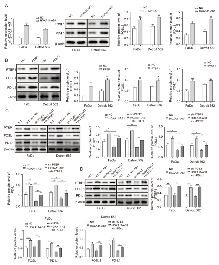
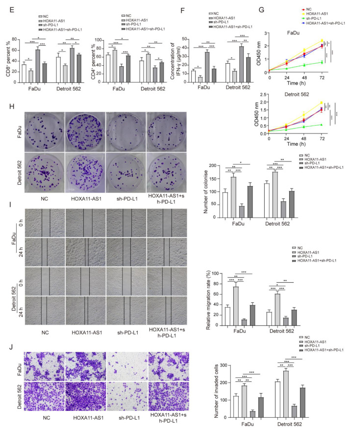
HOXA11-AS1 promoted PD-L1 expression by upregulating FOSL1 levels through PTBP1, thereby facilitating immune escape, growth, and metastasis of HSCC cells. FaDu and Detroit cells were transfected with pcDNA3.1-HOXA11-AS1 (HOXA11-AS1), pcDNA3.1-PTBP1 (PTBP1), sh-PTBP1, sh-PD-L1, or a combination of HOXA11-AS1 + sh-PTBP1 and HOXA11-AS1 + sh-PD-L1: (A) HOXA11-AS1, FOSL1, and PD-L1 levels were measured in FaDu and Detroit 562 cells treated with HOXA11-AS1 plasmid. (B) Relative expression of PTBP1, FOSL1, and PD-L1 was evaluated after overexpressing PTBP1. (C) Relative levels of PTBP1, FOSL1, and PD-L1 in HOXA11-AS1, shPTBP1, or HOXA11-AS1 + shPTBP1 groups. FaDu and Detroit cells were transfected with HOXA11-AS1, shPD-L1, or a combination of HOXA11-AS1 + shPD-L1: (D) Relative expression of PTBP1, FOSL1, and PD-L1 in cells transfected with HOXA11-AS1, sh-PD-L1, or HOXA11-AS1 + sh-PD-L1. Then, (E) the percentage of CD8+ and CD4+ T cells and (F) the concentration of IFN-γ were measured by flow cytometry and ELISA, respectively, and (G–J) the viability, colony formation, migration, and invasion of treated HSCC cells were analyzed by CCK-8, colony formation, wound healing, and transwell assays. Representative images (200×) are shown. * p < 0.05, ** p < 0.01, *** p < 0.001. Original Blots see Supplementary File Figure S1.


