Figure 4.
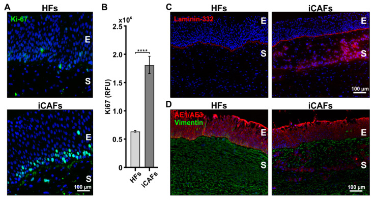
The presence of iCAFs in the stroma of 3D vesical models induces the proliferation and infiltration of urothelial cells. (A) Representative fluorescent images of the HFs-derived and iCAFs-derived models stained for Ki67 (green). Nuclei were counterstained with Hoechst (blue). (B) Corresponding quantification in relative fluorescence units (RFU) of the Ki67 signal. N = 4 independent biological replicates. (C) Representative fluorescent images of the HFs-derived and iCAFs-derived models of laminin-332. (D) Representative fluorescent images of vimentin- and epithelium-specific cytokeratin marker AE1/AE3 in the HFs-derived and iCAFs-derived constructs. The images show infiltration of epithelial cells within the stroma in the iCAFs-derived stroma. E = epithelium; S = stroma. The data are represented as mean ± SEM. **** p < 0.0001.

