Figure 5.
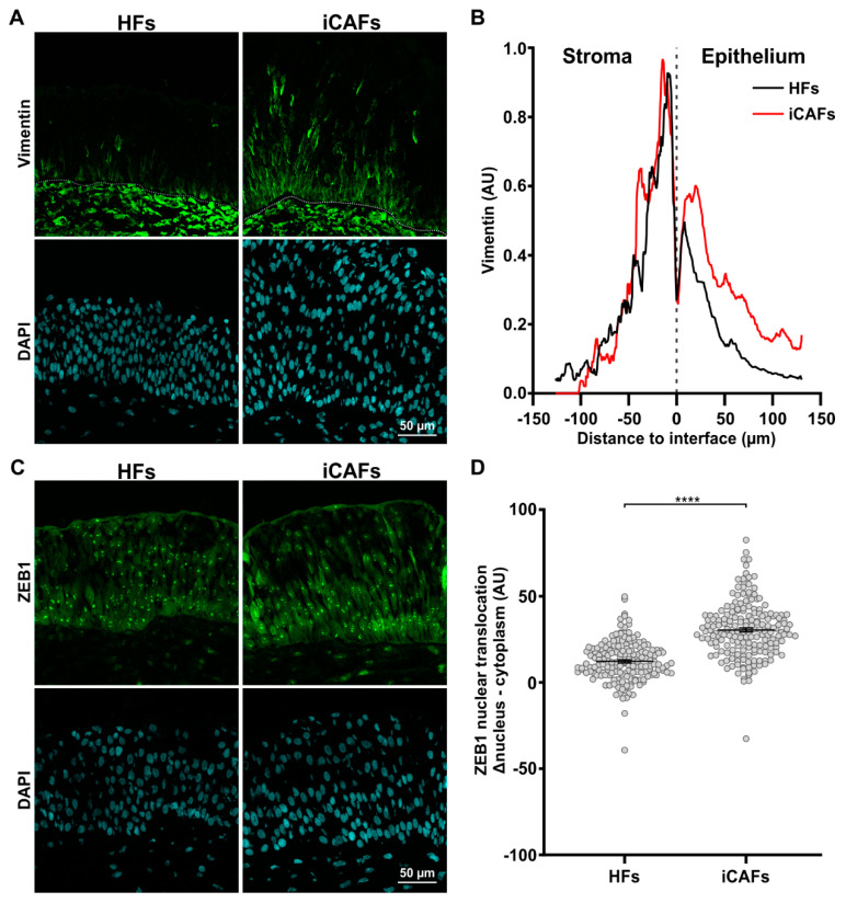
The iCAFs-derived ECM promotes an EMT state in urothelial cells. (A) Representative confocal images of the urothelium seeded on HFs-derived and iCAFs-derived stroma and stained for vimentin. (B) Corresponding quantification of the vimentin signal across the urothelium. The basal lamina was set as the origin and positive values go toward the top of the urothelium. (C) Representative confocal images of the urothelium seeded on HFs-derived and iCAFs-derived stroma and stained for ZEB1. (D) Corresponding quantification of ZEB1 signal by measuring the difference between nuclear expression and cytoplasm expression in urothelial cells. Data are presented as scatter plots (N = 3 per condition, 15 images per condition). Nuclei were counterstained with DAPI. The data are represented as mean ± SEM. **** p ≤ 0.0001.

