Figure 1.
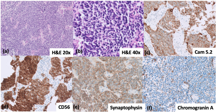
(a–f) Immunohistochemical patterns for the diagnosis of small-cell lung cancer. (a,b) H&E image of SCLC at 20× and 40× magnification, respectively. (c) CAM 5.2 with a dot-like pattern of cytoplasmic positivity; (d) CD56, strong positivity; (e) Synaptophysin, diffusely positive; (f) Chromogranin A, dot-like positivity in cytoplasm, pathognomonic of SCLC. (b–f) taken at 40× magnification.

