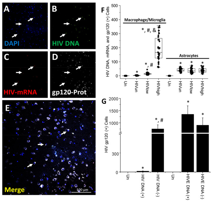
Figure 3.
HIV-gp120 protein is expressed in myeloid and glial viral reservoirs and surrounding uninfected cells: potential bystander toxicity under the current ART era. (A–E) Representative confocal images showing (A) DAPI staining, (B) HIV DNA, (C) Macrophage/Microglia marker Iba-1 protein, (D) HIV-gp120 protein (arrows denote triple-positive cells), and (E) corresponds to the merging of all colors. (F) Quantified Macrophage/Microglia and astrocyte cells with integrated HIV DNA, producing HIV-mRNA and HIV-gp120 according to the viral replication status, including undetectable, low, and high replication (* p ≤ 0.005 compared to uninfected conditions, # p ≤ 0.005 compared to HIVun conditions, and & p ≤ 0.005 compared to HIVun or HIVlow conditions; n = 34 tissues analyzed and 21 tissues compared to uninfected tissues, Un and Alz, n = 8 different tissues; each point represents 3–5 different areas per tissue analyzed). HIVE total numbers were 1364 ± 279 cells, 876 ± 458 for macrophages, and 624 ± 287 for astrocytes under HIVE conditions (data not plotted). (G) Quantification of cells positive for HIV-gp120 detected in uninfected cells (HIV DNA (−)) surrounding the clusters containing integrated HIV DNA (HIV DNA (+)) corresponded to 750.61 ± 164.85 cells, suggesting bystander damage within the CNS (* p ≤ 0.005 compared to uninfected conditions and # p ≤ 0.005 compared to HIV DNA (+) cells). In contrast, only 10.45 ± 3.65 cells with integrated HIV DNA accumulated HIV-gp120 protein. In HIVE conditions, cells with integrated HIV DNA (HIVE-DNA (+)) were 1364 ± 349 cells, and uninfected cells (HIVE-DNA (−)) were 901 ± 302 cells; thus, local numbers of cells containing HIV-gp120 could be comparable to HIVE conditions but with a significantly lower expression or accumulation as well as being extremely localized. Bar: 100 µm.

