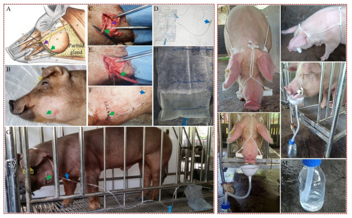
Figure 2.
Development of a unilateral parotid duct cannulation-based (UPDCB) surgical method (left panel) and a bridle-like device-based (BLDB) nonsurgical method (right panel) for efficient collection of saliva from hNGF-TG pigs. (A) An anatomical drawing of the pig face showing the position of parotid gland and its duct extending to the oral cavity. (B) A pig that is ready (under anesthesia and with its facial hair shaved) for unilateral parotid duct cannulation surgery. (C) Exposure of the parotid duct after facial surgery. (D) A medical urethral catheter and a medical drainage bag used for the parotid duct cannulation. (E) Cannulation of the parotid duct with a urethral catheter. (F) Suture of the facial wound after the parotid duct cannulation surgery. (G) Collection of parotid saliva from a pig by the UPDCB method. (H) The TG pigs’ parotid saliva collected into a drainage bag by the UPDCB method. Green, purple, and blue arrows point to the position of parotid duct, facial nerve, and urethral catheter connector, respectively. The front (I) and lateral (J) view of a TG pig wearing a bridle-like device consisting of a headstall and a bit, which were made of a rope and a plastic pipe (about 3 cm in diameter), respectively. The bit is attached by the headstall, which keeps the bit in place in the mouth to make the mouths of TG pigs slightly open. The front (K) and lateral (L) view of a TG pig wearing a bridle-like device attaching with a simple saliva collection device, which is composed of a funnel and a bottle. (M) The TG pigs’ oral saliva collected into a bottle by the BLDB method. Collection of TG pigs’ oral saliva by the BLDB nonsurgical method also was shown in Supplementary Video S1.

