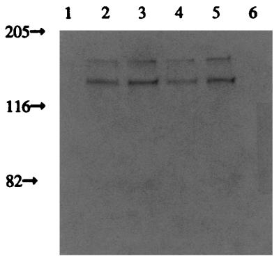
FIG. 3.
Sln1ΔTMD1p subcellular localization. The SLN1ΔTMD1-expressing strain was examined for Sln1p subcellular distribution by immunoblot analysis using anti-Sln1p antisera. Cells were grown to the mid-logarithmic phase of growth, harvested, and lysed, and membrane-associated and cytosolic proteins separated. The total membrane fraction was used to make a crude plasma membrane preparation by differential centrifugation. The purity of the plasma membrane fraction was tested by the ability of plasma membrane H+-ATPase activity to be specifically inhibited by vanadate (see Materials and Methods). The remaining membranes were termed the microsomal fraction. Lanes 1 to 3, high-copy-number vector; lanes 4 to 6, low-copy-number vector. Lanes 1 and 6, total cytosolic protein; lanes 2 and 5, plasma membrane fraction; lanes 3 and 4, microsomal fraction. Sizes here and in Fig. 5 to 7 are in kilodaltons.

