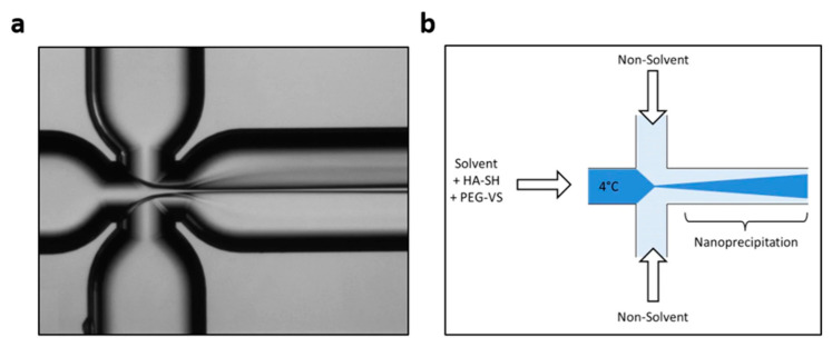
Figure 5.
Schematic illustration of the microfluidic experimental setup used by Tammaro et al. (a) Optical Fluorescence Microscopy Image of Flow-Focusing pattern; (b) Qualitative Illustration of crosslinking strategies. Reprinted from an open-access source [52].

