Figure 1.
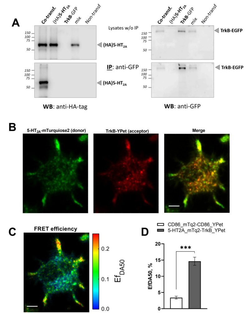
Interaction between receptors 5-HT2A and TrkB in neuroblastoma N1E-115 cells. (A) Co-immunoprecipitation of recombinant HA-tagged 5-HT2A and GFP (mEGFP)-tagged TrkB. Using a mixture of cells expressing each individual protein (mix) or cells that co-expressed both proteins (co-transfection), we performed immunoprecipitation (IP) with an anti-GFP antibody, followed by Western blot (WB) analysis with anti-GFP (right) and anti-HA (left) antibodies. Top: Expression of proteins before IP (lysate w/o IP). Bottom: Expression of proteins after IP. Results are representative of at least three independent experiments. (B–D) Specific interaction between 5-HT2A-mTurquoise2 and TrkB-YPet. Cells co-expressing 5-HT2A-mTurquoise2 and TrkB-YPet were analyzed using the lux-FRET method after confocal microscopy. (B) Distributions of 5-HT2A-mTurquoise2 (donor) and TrkB-YPet (acceptor), and merged images quantified by linear unmixing of the fluorescence emission spectra. Scale bar, 5 µm. (C) Apparent FRET efficiency (EfDA). A representative cell is shown. (D) Quantification of FRET efficiency (EfDA) between 5-HT2A-mTurquoise2 and TrkB-YPet. *** p < 0.001 (one-way ANOVA).

