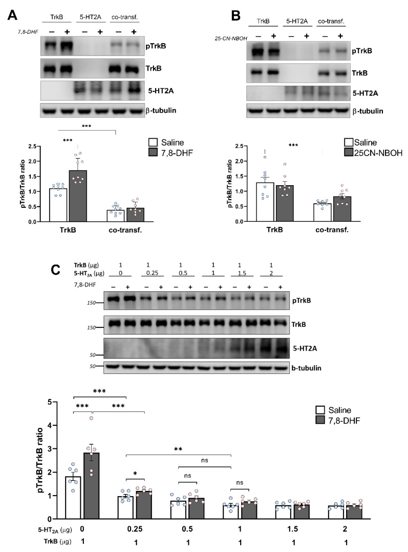
Figure 4.
Heterodimerization affects TrkB phosphorylation. (A,B) Analysis of TrkB phosphorylation. Neuroblastoma N1E-115 cells expressing either HA-tagged 5-HT2A (1 µg), GFP-tagged TrkB (1 µg), or co-expressing equal amounts of both receptors were treated for 30 min with either (A) the 5-HT2A receptor agonist 25CN-NBOH (1 μM) or (B) the TrkB agonist 7,8-DHF (500 nM). Cells were lysed, followed by SDS-PAGE and Western blot (WB) analysis using antibodies against either total TrkB or phosphorylated TrkB. The 5-HT2A receptor was detected in parallel. In WB, ß-tubulin was used as a loading control. Representative Western blots are shown. Lower panels show the quantification of TrkB phosphorylation, which was performed by densitometry and calculated as the ratio of total TrkB expression to the TrkB phosphorylation signal after adjustment for the general expression level. Bars show means ± SEM (n ≤ 8). *** p < 0.001 (two-way ANOVA). (C) Heterodimerization affects agonist-mediated TrkB phosphorylation. Neuroblastoma cells were co-transfected with 1 μg of cDNA encoding the GFP-tagged TrkB receptor, together with increasing concentrations of HA-tagged 5-HT2A receptor. Cells were treated for 30 min with either 500 nM 7,8-DHF or vehicle, followed by WB analysis. Lower panel: Quantification of TrkB phosphorylation. Bars show means ± SEM (n = 6). ns: not significant, * p < 0.05, ** p < 0.01, *** p < 0.001 (two-way ANOVA).

