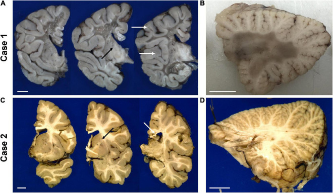
FIGURE 4.
Representative gross photographs of coronal sections of the forebrain (A,C) and sagittal sections of the cerebellum (B,D) of the untreated case 1 (top row) and transplanted case 2 (bottom row). The white arrows in panel (A) indicate examples of residual white matter between preserved cortical ribbon and end-stage white matter. The white arrow in panel (C) points to thinned posterior corpus callosum. The black arrow in panels (A,C) indicate the internal capsule. The scale bar is set to 1 cm for each panel.

