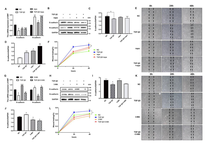
Figure 4.
Autophagy regulated the EMT process in HLE cells. (A) The mRNA levels of the EMT markers E-cadherin and N-cadherin in TGF-β2 and rapa (200 nM) co-treated cells. Mean ± SD, n = 3, * p < 0.05, ** p < 0.01, *** p < 0.001, **** p < 0.0001. (B) The protein levels of E-cadherin and N-cadherin in TGF-β2 (10 ng/mL) and rapa (200 nM) co-treated HLE cells. (C,D) Quantitative Western blot analysis (normalized to GAPDH). Mean ± SD, n = 3, * p < 0.05, ** p < 0.01. (E) Wound healing assays were conducted on HLE cells treated with TGF-β2 and with or without rapa; the images were photographed at 0, 24, and 48 h after scratching. (F) Quantitative wound healing area relative to 0 h. (G) The mRNA levels of E-cadherin and N-cadherin in TGF-β2 and 3-MA (10 mM) co-treated cells. (H) The protein levels of E-cadherin and N-cadherin. (I,J) Quantitative Western blot analysis (normalized to GAPDH). Mean ± SD, n = 3. * p < 0.05, ** p < 0.01. (K) Wound healing assays of TGF-β2 and 3-MA co-treated cells. (L) Graph shows the percentage of the wound healing area (normalized to 0 h).

