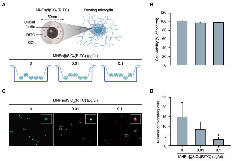
Figure 1.
Reduction in migratory activity of MNPs@SiO2(RITC)-treated BV2 cells. (A) Schematic representation of the invasion assay performed with MNPs@SiO2(RITC)-treated BV2 cells. Non-treated control BV2 cells, 0.01 µg/µL MNPs@SiO2(RITC)-treated BV2 cells, and 0.1 µg/µL MNPs@SiO2(RITC)-treated BV2 cells were used. (B) Viability of MNPs@SiO2(RITC)-treated BV2 cells for 12 h. (C) Invasion assay of MNPs@SiO2(RITC)-treated BV2 cells. The nuclei of the cells were stained using Hoechst 33342, and the fluorescence was expressed as cyan. Fluorescence of MNPs@SiO2(RITC) distribution was expressed as red. The number of nuclei was counted in five randomly selected fields. Scale bar = 50 μm. Scale bar of cropped image = 5 μm. (D) Number of migrating cells. Data represent means ± standard deviation derived from three independent experiments. * p < 0.05 vs. control.

