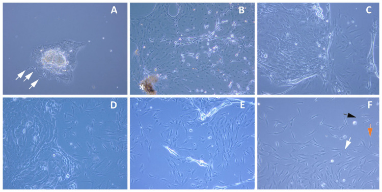
Figure 1.
Expansion of the limbal stem cells in culture. Representative images of the limbal stem cells in the primary culture: initiation (A) and at confluence (B,C), passage P1 (D), passage P2 (E) and passage P3 (F). Epithelial cells (round morphology) and stromal/progenitor cells (spindle morphology, indicated with white arrows) derived from the limbal explant (A). Gradual increase in limbal stromal cells population and simultaneous fading of limbal epithelial cells (C–E). Pure population of the limbal stromal cells obtained in passage P3 including dendritic cells ((F); black), undifferentiated myofibroblastic cells ((F); orange), and quiescent fibroblastic cells ((F); white); * Limbal Explant.

