Figure 1.
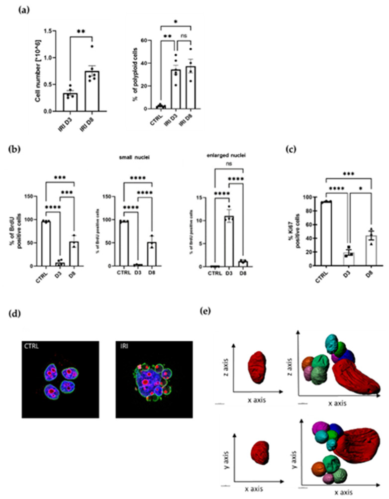
Therapy-induced senescence is associated with increased polyploidization and followed by resumption of proliferation. (a) MCF-7 breast cancer cells counted at day three (D3) and day eight (D8) after 24 h treatment with IRI (left panel); quantification of polyploid cells assessed with flow cytometry at day three (D3) and day eight (D8), >4c DNA cells were considered as polyploid (right panel); mean of at least three independent experiments ± SEM; statistical significance: * 0.01 < p < 0.05, ** 0.001 < p < 0.01. (b) Quantification of BrdU-positive MCF-7 cells; mean of at least three independent experiments ± SEM; statistical significance: * 0.01 < p < 0.05, ** 0.001 < p < 0.01, *** p < 0.001, **** p < 0.0001 (c) Quantification of Ki67-stained MCF-7 cells at day three (D3) and day eight (D8) mean of at least three independent experiments ± SEM; statistical significance: * 0.01 < p < 0.05, *** p < 0.001, **** p < 0.0001. (d) Representative images of control and treated cells stained with lamin a/c (green) and Ki67 (red), nuclei stained with Hoechst 33342 (blue). (e) 3D reconstruction of the serial block-face scanning electron microscopy (SBEM) sections. Representative reconstruction of control (on the left) and IRI-treated MCF-7 cells at D8 (on the right); scale bar = 50 µM.

