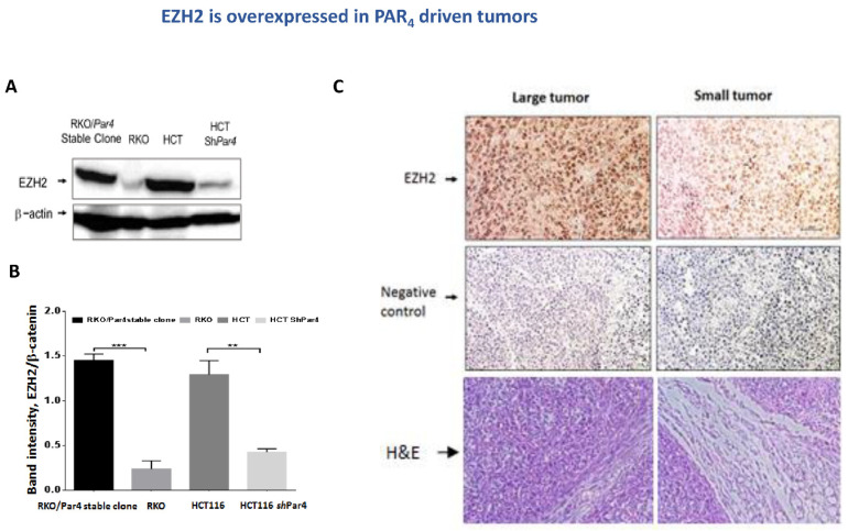
Figure 4.
EZH2 is overexpressed in PAR4-driven tumors. (A) Proteins were extracted from tumor lysates: wt RKO, RKO/Par4 clone, wt HCT-116, and shRNA-Par4-HCT116. Western blot analysis of the lysates was carried out. EZH2 was detected by anti-EZH2 antibodies (1:500) and β-actin by anti-β-actin antibodies (1:1000) as a control for protein loading. Western blot results that are representative of the assays performed three times are shown. (B) Quantification of bands using Image J software, normalized to β-Actin. (C) IHC of EZH2 in PAR4-driven tumors. Representative sections of mouse-generated PAR4-driven tumors. IHC staining, using anti-EZH2 (1:50 dilution) antibodies. All images were acquired using a Nikon light microscope at magnifications of 10× and 20×. Scale bars 50 µm. EZH2 is abundantly expressed in the large tumors (of high PAR4 (RKO/Par4a cells)). Very little to almost no EZH2 was detected in the small-appearing tumors (e.g., RKO). As controls for the IHC staining, tissue sections were processed in a similar fashion, but without primary antibodies.

