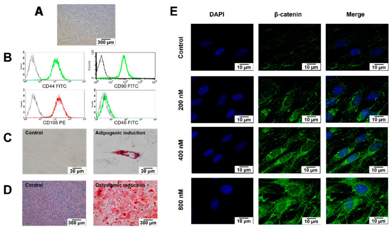
Figure 1.
Characterisation of the cells isolated from dental pulp tissues. (A) Morphological observation of human dental pulp stem cells (hDPSCs) using phase-contrast microscopy. Scale bars: 300 µm. (B) Evaluation of stem cell surface markers using flow cytometry. (C) Multi-lineage differentiation potential toward adipogenic (D) and osteogenic lineage. Scale bars: 30 and 300 µm. Intracellular lipid accumulation was stained by Oil Red O staining. Calcium accumulation was stained using Alizarin Red S (ARS) staining. (E) Representative images of immunofluorescence staining of β-catenin (stained in green) in hDPSCs; nuclei were counterstained with DAPI (shown in blue). White arrows indicate the increased nuclear translocation of β-catenin. Scale bars: 10 µm.

