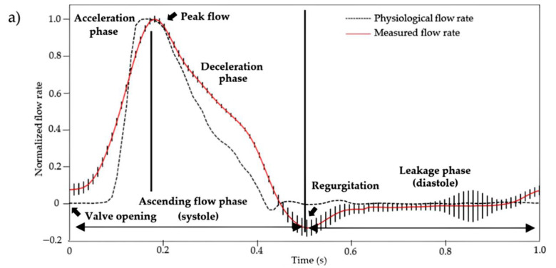
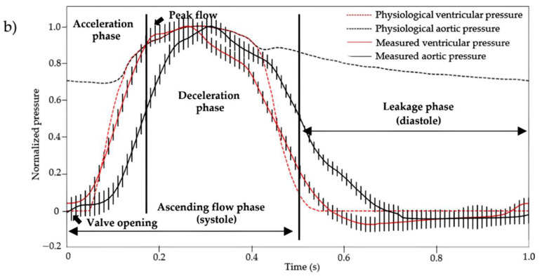
Figure 7.
(a) Average measured flow rate and (b) pressures (aortic and ventricular) normalized profiles (n = 12 cardiac cycles) at 60 BPM. These were obtained using a custom-made cardiac bioreactor on three aortic valves scaffolds (measured data curves) and compared to the physiological profiles (physiological value curves) [53]. The vertical bars represent the standard deviation at each acquisition time (100 Hz) of the average profiles of the three aortic valve scaffolds.


