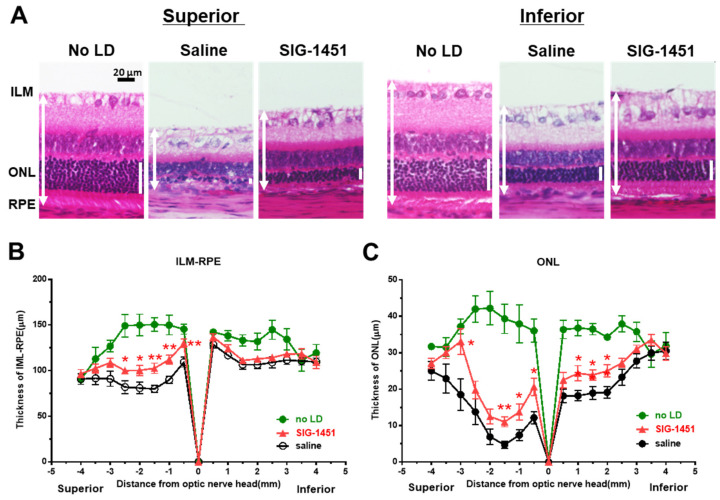
Figure 2.
Histological examination of retinas 8 d after light damage. Typical microphotographs of the superior and inferior parts of the retina (A). ILM-RPE (B) and ONL (C) thicknesses in saline- and SIG−1451-treated rats. Data are represented in terms of the mean ± SE values (saline: n = 14, SIG−1451: n = 16, unpaired t-test; *, ** p < 0.05, 0.01).

