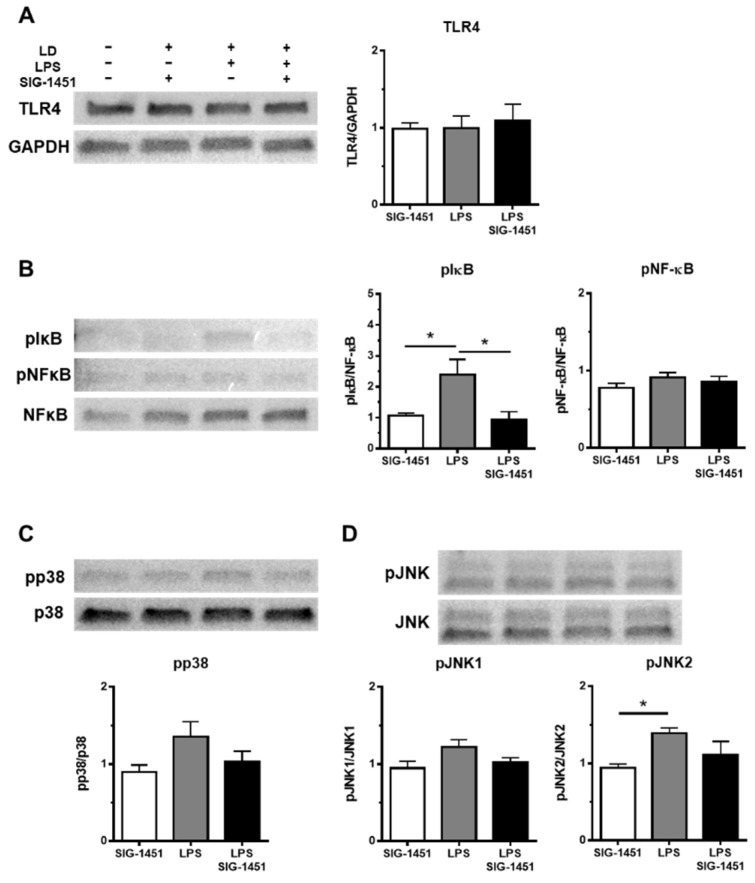
Figure 6.
Western blot analysis of inflammatory response-related proteins in rMC-1 cells. Total cell lysates were prepared 3 h after the LPS stimulation and used for the Western blot analysis. The levels of TLR4 were not changed between groups (A). The phosphorylation of I-κB, but not NF-κB, was increased by LPS stimulation and blocked by the addition of SIG-1451 (B). Phosphorylated p38 levels tended to increase upon LPS stimulation (C). Phosphorylated JNK level, particularly pJNK2, was significantly increased by LPS stimulation (D). Data are represented in terms of the mean ± SE values (n = 5, Tukey’s multiple comparisons test; * p < 0.05).

