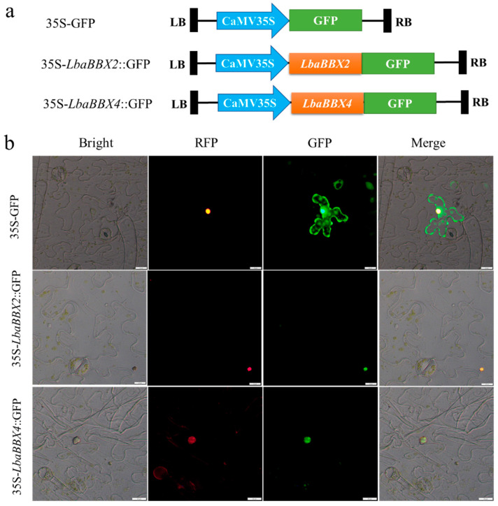
Figure 10.
Subcellular localization of LbaBBX4 protein. (a) Schematic diagram of the 35S-GFP, 35S-LbaBBX2::GFP, and 35S-LbaBBX4::GFP fusion protein constructs used for transient expression. (b) The LbaBBX2-GFP and LbaBBX4-GFP fusion proteins were transiently expressed in N. benthamiana leaves and observed by fluorescence microscopy 48 h later. The 35S-GFP was used as positive control. From left to right, bright field, red fluorescent protein (RFP) (nuclear localization signal (NLS)-RFP), green fluorescent protein (GFP), and merge image of RFP and GFP image. Scale bars =20 μm.

