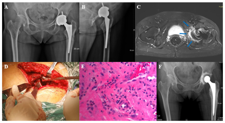
Figure 4.
Typical case 2. Female, 68 years old, left total hip arthroplasty. (A,B): X-ray before stage I resection surgery; (C): MARS MRI showed signs of acetabular bone destruction and bone edema (at the arrow); (D): intraoperative exploration revealed occult lesions in the acetabulum; (E): histopathology of the acetabulum suggests infection; (F): X-ray after stage I resection surgery.

