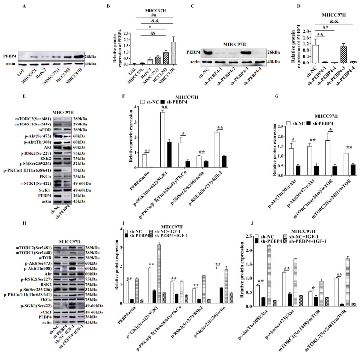
Figure 1.
The effect of PEBP4 knockdown on the mTORCs-mediated signaling pathways. (A) Western blot (WB) was performed using different HCC and immortalized normal hepatocytes as indicated. (B) Scan densitometric ratio of PEBP4 to actin. (C,D). MHCC97H cells were infected with sh-NC and different PEBP4 shRNA1-4. Cell extracts were blotted (C) and quantified by scan densitometry (D). (E) Extracts of MHCC97H cells with sh-PEBP4 and sh-NC were blotted with antibodies. (F,G) Relative scan densitometric units were determined by the ratio of phospho-proteins to their cognate total proteins or actin for (E). (H) MHCC97H cells with sh-PEBP4 or sh-NC were treated with or without IGF-1 (100 µg/mL) for 30 min and WB was performed using antibodies. (I,J) Relative scan densitometric units were determined for H. All graphs represent averages of three independent experiments (mean ± SEM). * p < 0.05, ** p < 0.01, ## p < 0.01, && p < 0.01, $$ p < 0.01.

