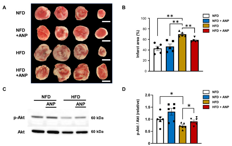
Figure 4.
ANP treatment improved IRI and insulin sensitivity in HFD hearts. (A) Micrograph showing representative triphenyltetrazolium chloride (TTC) staining of cardiac sections obtained from NFD, NFD + ANP, HFD, and HFD + ANP groups after ischemia–reperfusion. Bars = 3 mm. (B) Effects on the quantitated cumulative infarct area size in NFD, NFD + ANP, HFD, and HFD + ANP hearts (n = 5 each). (C) Representative immunoblots for phospho- and total Akt from cardiac lysates after ischemia–reperfusion. (D) Densitometric quantitation normalized to the level of p-Akt/total Akt expression in NFD hearts after IRI is shown (n = 6 each). * p < 0.05 and ** p < 0.01 versus HFD.

