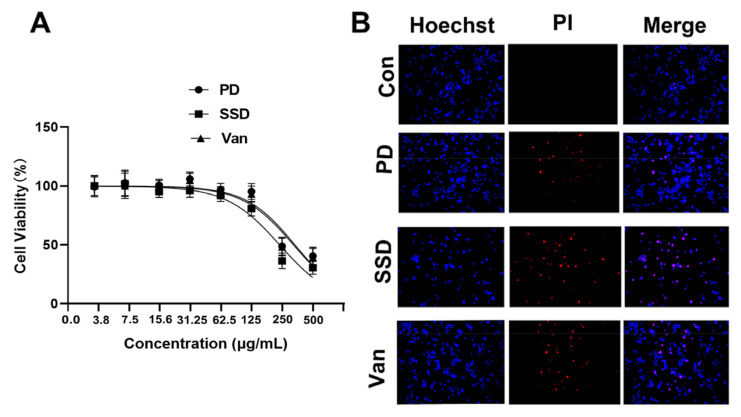
Figure 1.
The cytotoxicity of the PD, SSD, and Van on human keratinocytes (HaCaT). (A) HaCaT cells were treated with the indicated concentration of the PD, SSD, and Van for 24 h. Cell viability was assessed by MTT assay. (B) HaCaT cells were treated with 125 µg/mL of the PD, SSD, and Van for 24 h. Cell apoptosis was assessed by Hoechst/PI staining. The scale bar is 200 μm.

