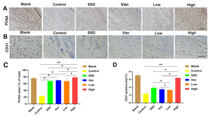
Figure 5.
Immunohistochemical analysis of cutaneous wound sections at day 14 post-wound healing/infection. (A) Analysis of proliferating cell nuclear antigen (PCNA) + cells in wounds at day 14 post-infection. Representative images of PCNA+ cells in wounds treated with Pd, SSD, Van, or the blank hydrogel. (B) Analysis of CD31 in wounds at day 14 post-infection. (C) Corresponding quantification of PCNA+ cells expressed as a percentage of total cell counts. (D) Statistical analysis of CD31 expressed at the wound site in different treatment groups on day 14. All data are presented as mean ± standard deviation. Significant differences are indicated with asterisks (* p < 0.05, ** p < 0.01, n = 3. (magnifications, ×200).

