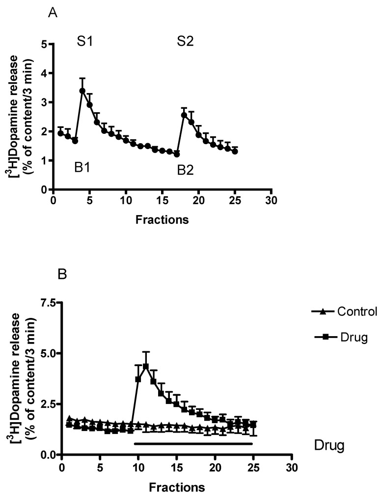
Figure 2.
(A) The time-course of [3H]dopamine release measured from rat striatum. Filled circles indicate [3H]dopamine release at rest and in response to electrical stimulation. Striatal slices were prepared, loaded with [3H]dopamine and superfused with aerated Krebs-bicarbonate buffer. [3H]Dopamine release was induced by electrical stimulation (40 V, 2 Hz, 2 msec for 3 min) in fractions 4 (S1) and 18 (S2) and was expressed as a fractional rate, i.e., a percentage of the amount of [3H]dopamine in the tissue at the time of the release. The calculated ratio of the electrically stimulated fractional release S2 (2nd stimulation) over fractional release S1 (1st stimulation) (S2/S1) was 0.84 ± 0.04, representing a release of vesicular origin. The calculated ratio of resting fractional release B2 (fraction17) over fractional release B1 (fraction 3) (B2/B1) was 0.72 ± 0.05. When studied, drugs were added to the superfusion buffer between the 1st and 2nd electrical stimulations and maintained through the experiment, mean ± S.E.M., n = 6. (B) The time-course of non-vesicular [3H]dopamine release from rat striatum. Striatal slices were prepared, loaded with [3H]dopamine and superfused with aerated Krebs-bicarbonate buffer. The release of [3H]dopamine was expressed as a fractional rate expressed as percent of content released. [3H]Dopamine release was induced by the addition of drugs to the superfusion buffer and maintained through the experiment. In this experiment, (±)amphetamine as a drug was added in a concentration of 10 µmol/L from fraction 10 and the evoked non-vesicular [3H]dopamine release was 13.88 ± 3.00 percent of content released (calculated between fractions 10 and 25). The calculated [3H]dopamine release was 0.53 ± 0.12 percent of content released in control conditions. Student’s t-statistics for two-means, p < 0.01, mean ± S.E.M., n = 4-4.

