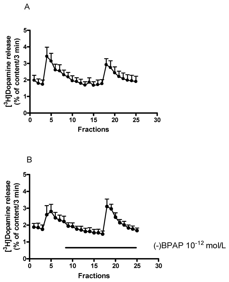
Figure 5.
(−)BPAP increased electrical stimulation-induced [3H]dopamine release from rat striatum. For the experimental procedure, see Figure 2A. When studied, (−)BPAP was added in a concentration of 10−12 mol/L to the superfusion buffer from fraction 8 and maintained throughout the experiment. (A) The release of [3H]dopamine from striatal slices, control experiments. Filled circles indicate [3H]dopamine release at rest and in response to electrical stimulation. The resting [3H]dopamine release (B2/B1) and the electrical stimulation-induced [3H]dopamine release (S2/S1) were 0.92 ± 0.05 and 0.74 ± 0.04 in control conditions. (B) Effect of (−)BPAP (10−12 mol/L) on [3H]dopamine release from rat striatum. Filled circles indicate [3H]dopamine release in the presence and absence of (−)BPAP. (−)BPAP was added to striatal slices from fraction 8 and maintained throughout the experiment. The resting [3H]dopamine release (B2/B1) and the electrical stimulation-induced [3H]dopamine release (S2/S1) were 0.96 ± 0.08 and 1.24 ± 0.13 in the presence of (−)BPAP. These values indicate that (−)BPAP in 10−12 mol/L concentration failed to influence the resting (p = 0.55) but increased the electrically induced [3H]dopamine release (p < 0.01). Student t-statistics for two-means, mean ± S.E.M., n = 7-6.

