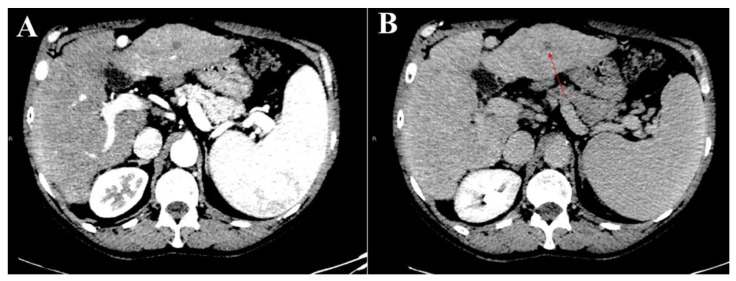
Figure 2.
Representative sets of CT images of an atypical liver nodule in segment three in the arterial (A) and the delayed (B) phases (red arrow) were presented to the readers in PDF format. In particular, the nodule was hypodense in both the arterial and the delayed phases, and its biopsy was considered feasible.

