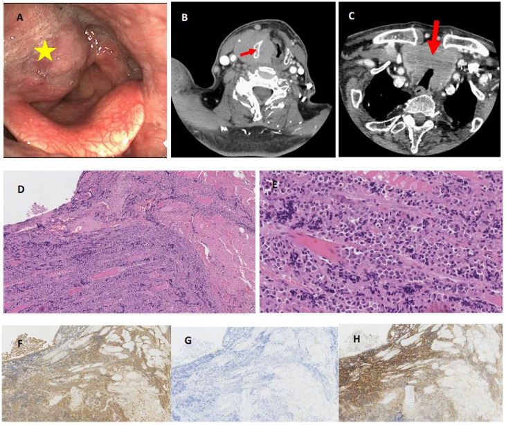
Figure 2.
Endoscopic view of bulky extramedullary plasmacytoma invading right side of the larynx and hypopharynx (yellow asterisk) (A). Neck CT scan of large tumour mass involving laryngeal structures at the glottic and supraglottic level, with extralaryngeal spread and thyroid cartilage sclerotization and partial destruction (red arrow) without increased tissue enhancement (B). The infiltration involves also both thyroid gland lobes with moderate narrowing of the trachea (red arrow) (C). Histological view of hematoxylin and eosin staining of laryngeal specimen with plasma cells infiltrating muscles x10 (D) x40 (E) and strong cytoplasmic kappa light chain positivity x10 (F) with complete absence of lambda light chain expression x10 (G) and immunohistochemical positivity for CD138 antigen x10 (H).

