Figure 1.

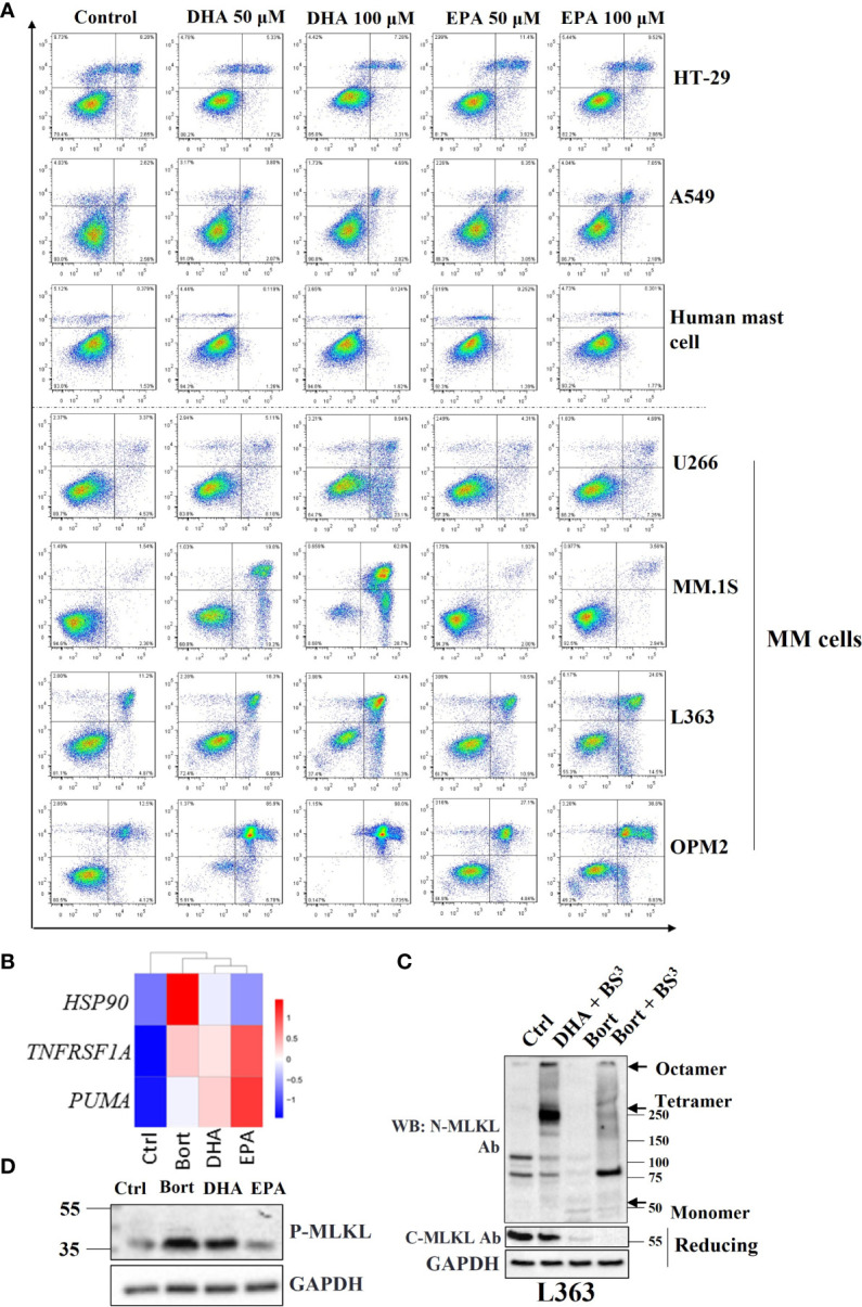
Necroptosis is involved in DHA/EPA and bortezomib-induced cell death in MM cells. (A) Cells were treated with 50 µM and 100 µM of DHA or EPA for 24 h. Cell death was determined by Annexin V and PI staining. (B) OPM2 cells were treated with 50 µM of DHA/EPA or 10 nM of bortezomib. RNA sequencing was performed. The expression levels of HSP90, TNFRSF1A and PUMA were shown in heatmap. (C) L363 cells were treated with DHA (50 µM) or bortezomib (500 nM) for 24 h and lysed after 1 h incubation with crosslinker BS3. Cell lysates were subjected to SDS-PAGE under non-reducing and reducing conditions and immunoblotted with indicated antibodies. Arrows indicate the oligomers of MLKL. (D) Cells were treated with 100 µM of DHA or EPA and 500 nM of bortezomib for 24 h and lysed with RIPA buffer. Whole cell lysates were subjected to western blotting with antibodies against p-MLKL and GAPDH.
