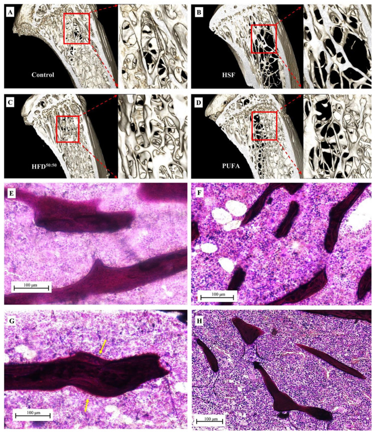
Figure 7.
3D models of the proximal tibia were created using 3D Slicer. (A) Control, (B) HSF-fed group showing evidence of trabecular bone thinning and bone loss, (C) HFD50:50 diet demonstrating thicker and denser trabeculae when compared with all other groups and (D) the PUFA-fed group shows increased porosity compared with the control-fed and HFD50:50-fed animals. Micrographs taken using light transmission microscopy of cancellous bone in the proximal tibia in the (E) control, (F) HSF, (G) HFD50:50 and (H) PUFA groups. Increased trabecular thickness was observed in the HFD50:50 group, with areas of osteogenesis (yellow arrows) observed on the trabecular surface. In contrast, the surfaces of bone appeared quiescent in all other experimental groups, with thinner trabeculae seen in the HSF and PUFA samples.

