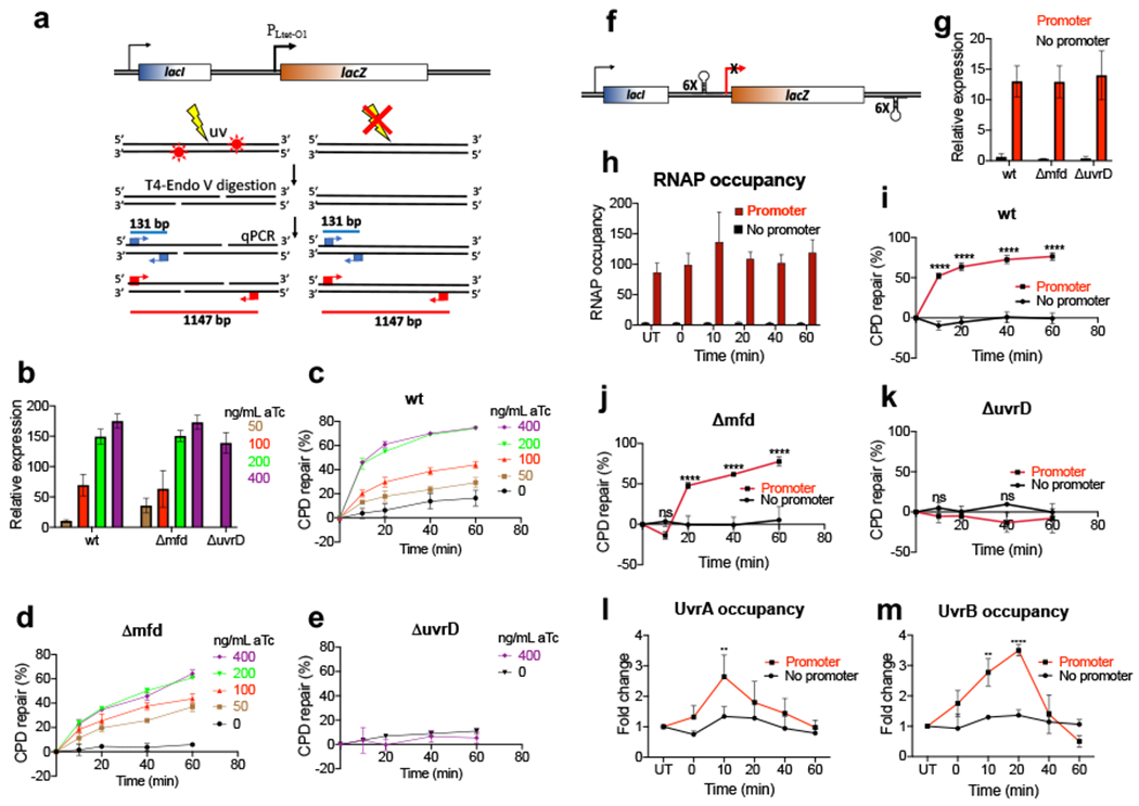
Fig. 5. Local transcription enables NER (independently of Mfd).

a, Schematic illustration of the semi-long-range (SLR)-qPCR assay to quantitate CPDs within the ROI (see Methods). Cells were exposed to UV followed by the isolation of genomic DNA (top). DNA was treated with T4endoV to convert CPDs to single strand breaks (SSBs) (middle). In the subsequent qPCR step the undamaged ROIs of 1147 bp (red) are successfully amplified, whereas SSBs abrogate PCR in damaged ROIs. Short fragments of 131 bp (blue) serve as a reference. Accumulating CPDs increase ΔCp of qPCR (bottom) allowing for an accurate CPD quantitation per 10 kb. b-e, The rate of local CPD repair as a function of promoter strength. b, Induction of chromosomal PLtet-O1-lacZ by the increasing concentration of anhydrotetracycline (aTc), as determined by RT-qPCR relative to a reference constitutive gene (cysG). Values are means ± SD (n = 3). c-e, Repair of CPDs within the PLtet-O1-lacZ ROI in wt (c), Δmfd (d), and ΔuvrD (e) cells. Transcription was induced by the indicated amounts of aTc as in (b) followed by UV irradiation (40 J/m2). Cells recovered in dark for the indicated time intervals followed by CPD quantitation as in (a). Values are the means ± SD (n = 3). f-k, Depriving a genomic locus of transcription abolishes its NER (irrespectively of Mfd) (see also Extended Data Fig. 11). f, Schematics of the lacZ insulator. Chromosomal lacZ, with or without its native promoter, was insulated from a possible upstream and downstream transcriptional readthrough with the intrinsic terminator cassettes. g, Expression of lacZ from the insulator upon IPTG induction, as determined by RT-qPCR. Values are means ± SD (n = 3). h, Occupancy of RNAP before (UT) and after UV irradiation, as determined by chromatin immunoprecipitation followed by qPCR (ChIP-qPCR). Cells were induced with IPTG followed by UV irradiation (40 J/m2) and recovery for the indicated time intervals. Values are means ± SD (n = 3). i-k, CPD repair within the insulator. Bulk of CPD repair in wt (i) and Δmfd (j) cells strictly depends on promoter-initiated transcription. No significant repair within the insulator with or without promoter was detected in ΔuvrD cells (k). Cells were induced with IPTG followed by UV irradiation (40 J/m2) and recovery in the dark for the indicated time intervals. CPD density was determined by SLR-qPCR as in (a) and used to calculate the percentage of repaired CPDs. Values are means ± SD (n=3), **P < 0.01, ****P < 0.0001 (Student’s t test; equal variance). l,m UvrAB recruitment to the UV-damaged DNA strictly depends on local transcription (see also Extended Data Fig. 11). Recruitment of UvrA (l) and UvrB (m) to the lacZ insulator (with or without promoter) was determined by ChIP-qPCR as in (h). Results are shown as a fold change in the occupancy of UvrAB within the insulator following UV irradiation. **P < 0.01, ****P < 0.0001 (Student’s t test; equal variance).
