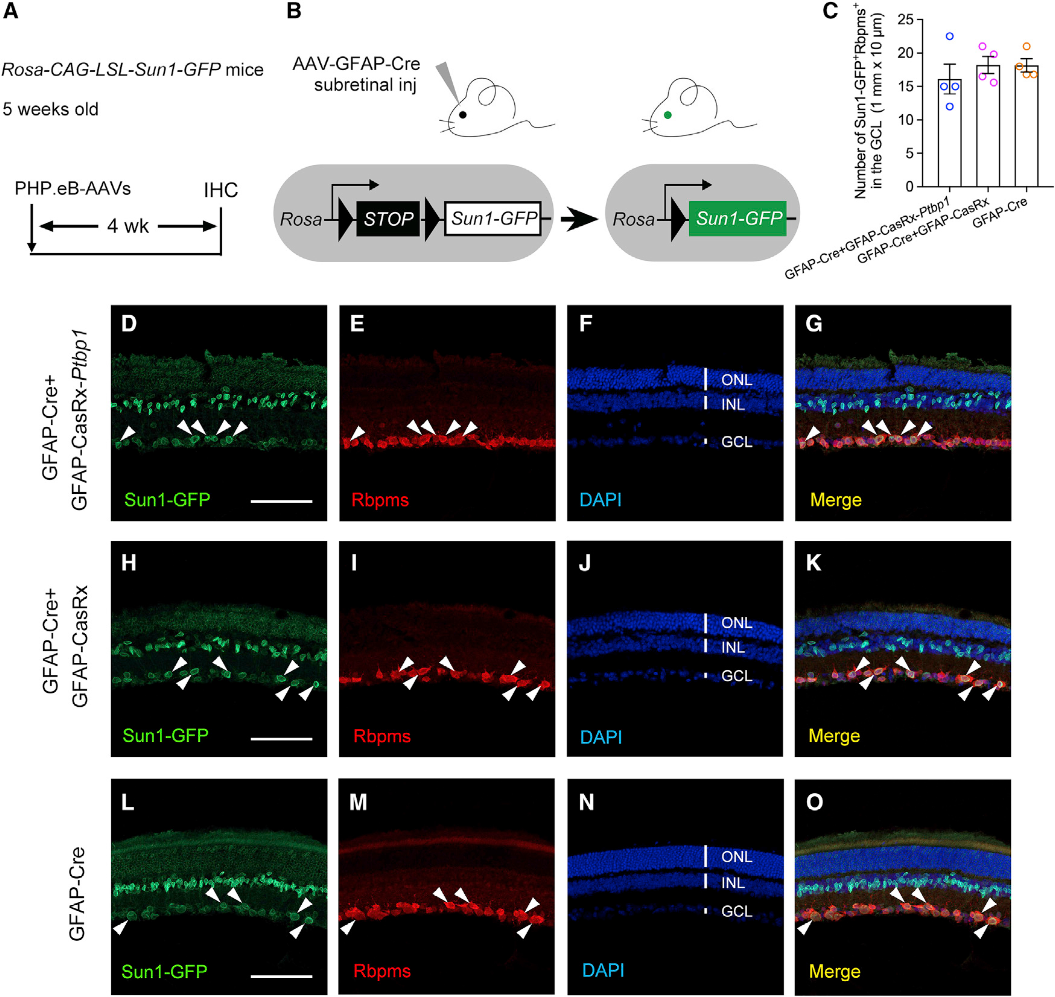
Figure 1. AAV-mediated Cre recombination labels endogenous RGCs and thus is unsuitable for examining MG-to-RGC conversion.

(A) Experimental design for testing MG-to-RGC conversion. Subretinal injection of PHP.eB-AAVs was performed in Rosa-CAG-LSL-Sun1-GFP reporter mice at 5 weeks of age, followed by immunohistochemistry analysis using the pan-RGC marker RBPMS at 4 weeks after AAV injection. IHC, immunohistochemistry.
(B) Schematic illustration showing that AAV-GFAP-Cre subretinal injection results in Sun1-GFP expression after Cre-dependent recombination in Sun1-GFP reporter mice.
(C) Quantification of Sun1-GFP+Rbpms+ cells in the ganglion cell layer (GCL) at 4 weeks after AAV injection. n = 4 retinas per group. Data are presented as the mean ± SEM. No significant difference was found between any two groups, p > 0.5, unpaired t test.
(D–G) Confocal images showing expression of Sun1-GFP and RBPMS immunohistochemistry after Ptbp1 knockdown in retinas receiving GFAP-Cre and GFAP-CasRx-Ptbp1. White arrowheads: Sun1-GFP-labeled cells were RBPMS positive in the GCL.
(H–K) Confocal images showing expression of Sun1-GFP and RBPMS immunohistochemistry in retinas receiving GFAP-Cre and GFAP-CasRx. White arrowheads: Sun1-GFP-labeled cells were RBPMS positive in the GCL.
(L–O) Confocal images showing expression of Sun1-GFP and RBPMS immunohistochemistry in retinas receiving GFAP-Cre only. White arrowheads: Sun1-GFP-labeled cells were RBPMS positive in the GCL.
(D–O) Scale bars, 100 μm. ONL, outer nuclear layer; INL, inner nuclear layer; GCL, ganglion cell layer. See also Figure S1.
