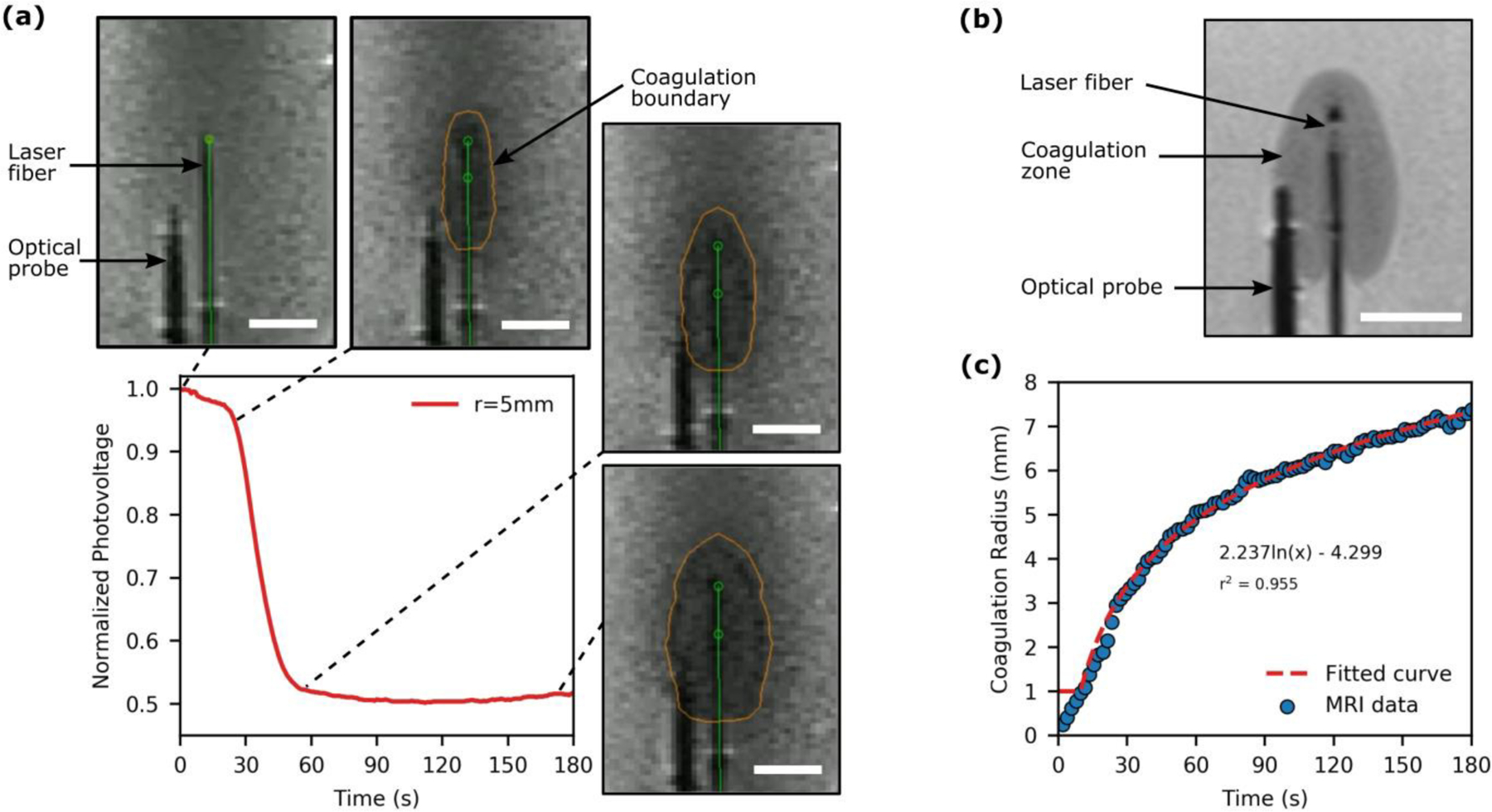
Fig. 4.

Analysis of laser-tissue interaction during FLA in a tissue-mimicking phantom under simultaneous MRI surveillance. (a) The acquired optical signal and corresponding MRI images (INTRA) for FLA with the ballistic probe positioned 5mm from the laser fiber. The optical signal decreases as the coagulation zone expands towards the probe with an inflection point observed once the coagulation zone reaches the probe. (b) A high-resolution MRI (POST) acquired immediately after FLA. (c) An example of the growth of the coagulation radius as a function of time which was described with a logarithmic function to facilitate temporal resolution with the acquired photovoltage data. [All scale bars are 10mm]
