Fig. 2.
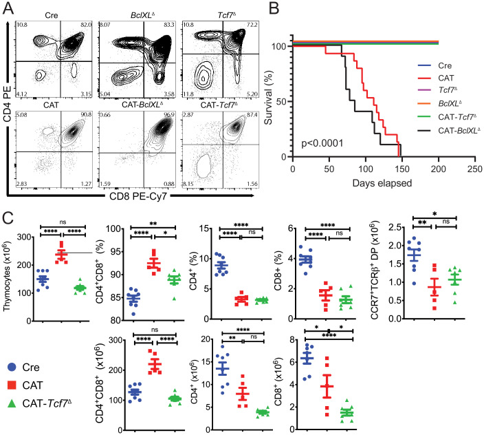
Ablation of Tcf7 rescues CAT lymphomas but a developmental block remains. (A) Representative flow cytometry contour plots of live thymocytes from 6- to 8-wk-old mice stained for CD4 and CD8 to show developmental progression. (B) Kaplan–Meier survival curve analysis for Cre (CD4-Cre, n = 10), CAT (CD4-Cre/Ctnnbex3fl/ex3fl, n = 15), Tcf7Δ (CD4-Cre/Tcf7fl/fl, n = 10), BclXLΔ (CD4-Cre/BclXLfl/fl, n = 10), and those with codeletions CAT-Tcf7Δ (CD4-Cre/Ctnnbfl/f/Tcf7fl/fl, n = 5) or CAT-BclXLΔ (CD4-Cre/Ctnnbfl/f/BclXlfl/fl, n = 10). (C) Flow cytometric histograms indicating the percentage (Top) and total number (Bottom) of thymocytes in the indicated late developmental stages in Cre (n = 8), CAT (n = 5), and CAT-Tcf7Δ (n = 7) mice; data are represented as the mean ± SEM, and statistical testing is depicted as two-sided, unpaired t tests; ns > 0.05. *P ≤ 0.05, **P ≤ 0.01, ****P ≤ 0.0001.

