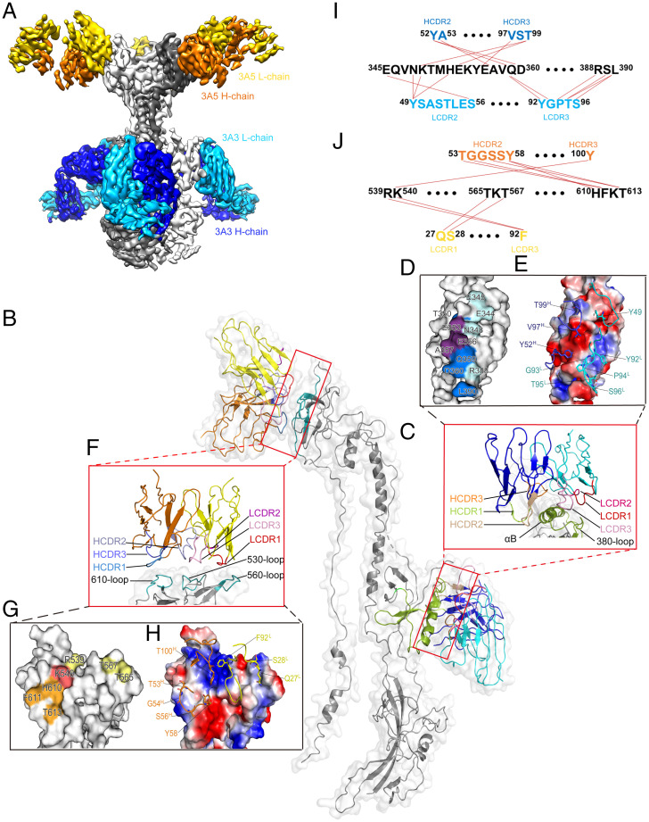
Fig. 5.
Structure determination of gB:3A3Fab:3A5Fab by cryo-EM. (A) Side view of the 3.9-Å cryo-EM structure of gB:3A3Fab:3A5Fab. gB, 3A3Fab, and 3A5Fab are colored gray, blue, and yellow, respectively. (B) Segmentation of monomeric gB binding with one 3A3Fab and one 3A5Fab. The key elements of D-II and D-IV of gB involved in antigen-antibody interactions are colored green and cyan, respectively. 3A3Fab and 3A5Fab are colored blue and yellow, respectively. (C-E) The interface of gB with 3A3Fab. The CDRs of the VH and VL and the gB’s αB-helix and 380 loop are shown differently (C). The residues involved in the 3A3 interactions are mapped on the gB surface, including E344, E345, N348, T350, E353, E356, A357, Q359, D360, R388, and L390 (D). The key residues localized at the VH and VL are labeled, including Y49L, Y92L, G93L, P94L, T95L, S96L, Y52H, V97H, and T99H (E). (F-H) The interface of gB with 3A5Fab. The CDRs of the VH and VL and the 530 loop, 560 loop, and 610 loop of gB are shown with different colors (F). The key residues involved in the 3A5 interactions are mapped on the gB surface, including R539, K540, T565, T567, H610, F611, and T613 (G). The key residues localized at the VH and VL are labeled, including Q27L, S28L, F92L, T53H, G54H, S56H, Y58H, and T100H (H). (I and J) Molecular interactions between gB and the VH and VL of mAbs 3A3 (I) and 3A5 (J).

