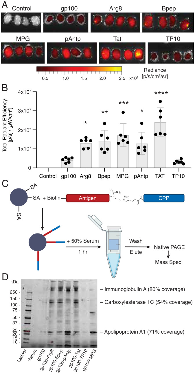
Fig. 5.
Antigen-CPP conjugates exhibit increased trafficking to dLNs and associate with serum proteins. (A) Whole-tissue fluorescence imaging of inguinal LNs 48 h after immunization with 25 nmol-labeled gp100 or gp100-CPPs with 25 µg of cyclic-di-GMP (n = 4 LNs per group, image shown at 1.5X)). (B) Quantification of total peptide fluorescence from each dLN, compared to control. ****P < 0.0001; ***P < 0.001; **P < 0.01; *P < 0.05; n.s., not significant by one-way ANOVA with Dunnett’s posttest. (C) Schematic of antigen pull-down experiment. (D) Native-PAGE analysis of proteins pulled down by gp100 or gp100-CPPs following incubation with mouse serum. Several bands identified by LC-MS/MS are indicated.

