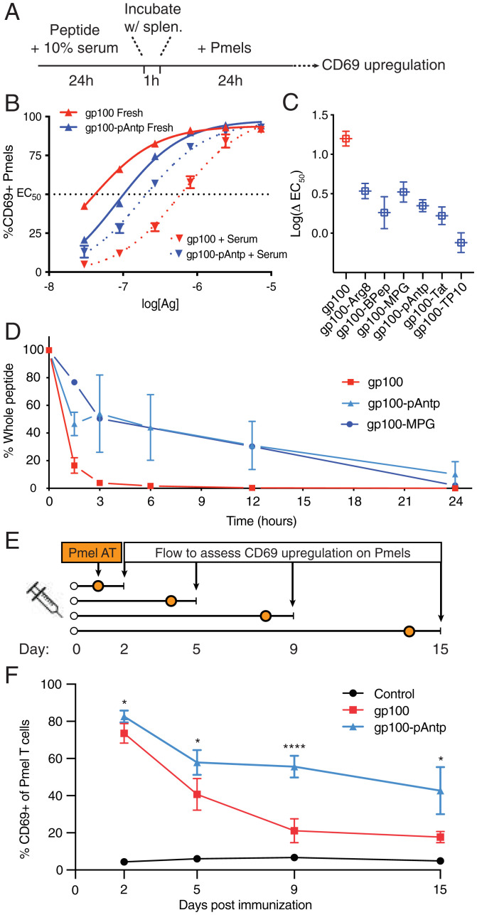
Fig. 6.
CPPs protect linked antigens from degradation in serum and prolong the duration of antigen presentation in dLNs. (A) Timeline of serum exposure T cell activation experiments. (B) Representative concentration dependence plots for CD69 up-regulation by pmel-1 T cells cocultured with splenocytes pulsed with the indicated concentration of gp100 or gp100-pAntp peptide with or without preincubation with serum. (C) Log fold-change in EC50 values for fresh vs. serum-treated peptides for each antigen construct. (D) Percentage of intact peptide remaining after incubation in 10% fresh mouse serum as analyzed by LC-QTOF-MS. (E) Timeline for experiments assessing the duration of antigen presentation following a single injection of gp100 or gp100-pAntp in C57BL/6 mice. AT, adoptive transfer. (F) Levels of available antigen as a function of time postimmunization in dLNs read out by pmel-1 T cell CD69 up-regulation (n = 5 animals per group). A two-way ANOVA with a Tukey posttest was used to compare gp100 and gp100-pAntp for each point, with: ****P < 0.0001, **P < 0.01, and *P < 0.05.

