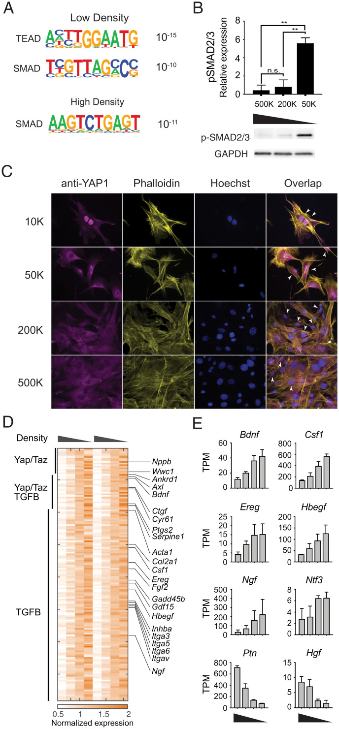
Fig. 3.
YAP1 and SMADs are activated in a density-dependent manner in fibroblasts. (A) Significantly enriched transcription factor binding motifs in genes induced at low or high density. (B) Phosphorylation of SMAD2/3 at different cell densities. (Upper) Quantification of phospho-Smad2/3 Western blot staining, normalized to GAPDH staining of the same sample, from three independent experiments. (Lower) Representative image of phospho-Smad2/3 and GAPDH Western blots. (C) Representative immunofluorescent images showing YAP1 localization in MEFs cultured at the specified cell densities overnight. Arrowheads denote examples of nuclear localization of YAP1. (D) Heatmap showing annotated targets of Hippo-YAP signaling and TGF-β signaling pathways at different cell densities. (E) Expression of representative growth factors significantly regulated by density (high to low density) **P < 0.01, t-test.

