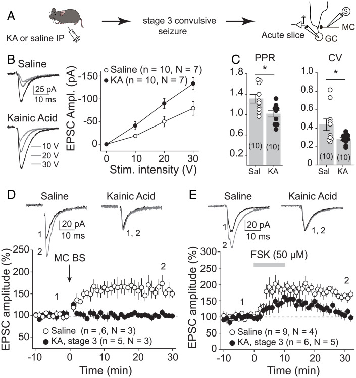
Fig. 3.
Initial convulsive seizures increased MC-GC synaptic strength. (A) Seizures were acutely induced using KA IP (20 mg/kg). Mice were killed humanely after reaching stage 3 of convulsive seizures, and MC-GC synaptic function was accessed in acute hippocampal slices. Saline-injected mice were used as control. (B) Representative traces and summary plot showing how input/output function was increased in KA-injected mice. EPSC amplitude (Ampl.) vs stimulus (Stim.) intensity is plotted. (C) PPR and CV were both significantly decreased in KA-treated mice as compared with saline-injected mice. Each number in parentheses represents the number of cells. (D and E) Representative traces (Top) and time course summary plots (Bottom) showing that LTP at MC-GC synapses induced by either MC BS (five pulses at 100 Hz, repeated 50 times every 0.5 s, D) or 50 μM forskolin (FSK, E) application was impaired in KA-injected mice. *P < 0.5. Data are presented as mean ± SEM.

