Fig. 5.
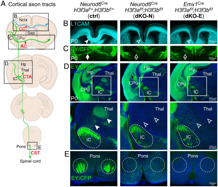
Defective axon tract development following H3f3a and H3f3b codeletion. (A) A schematic of major cortical axon tracts. (B) L1CAM (cyan) staining of P0 cortex. dKO-N and dKO-E were characterized by loss of white matter thickness and agenesis of the CC (arrowheads). (C–E) Cre-dependent fluorescent reporters expressed from the H3f3a and H3f3b floxed loci were detected by anti-EGFP immunostaining of EYFP and ECFP residues. (C) EY/CFP (green) reporter staining revealed failed midline crossing of the AC (arrows) and misrouting of AC axons to the hypothalamus in dKO-N and dKO-E. (D) In dKO-N and dKO-E, corticofugal axons reached internal capsule (IC), but corticothalamic tract axons (CTA, arrowheads) failed to innervate thalamus (Thal). (E) Analysis of the CST revealed an absence of axons that reached the level of the pons in dKO-N and dKO-E.

